Functional Cell-based Assays Verified & Compound Libraries
This unique platform is a powerful resource for researchers developing 3CLpro inhibitors and can streamline the development of safe and effective therapeutic treatments for COVID-19.
OTAVAchemicals and Montana Molecular have a complete solution for COVID-19 drug discovery that combines verified compound libraries with functional cell-based assays and drug development services.
The assay is 3CLglow, a live cell, fluorescent biosensor that reports target engagement and physiologically-relevant measurements of IC50 in living cells.
The OTAVA SARS-CoV-2 Main Protease Targeted Library contains 1017 compounds with predicted activity against SARS-CoV-2 3CL protease. The library has been designed with receptor-based virtual screening using crystal structure (PDB ID: 6LU7) of SARS-CoV-2 main protease. The structure of the enzyme active site was used to select compounds based on docking scores and intermolecular hydrogen bonds between key amino acid residues and the compound.
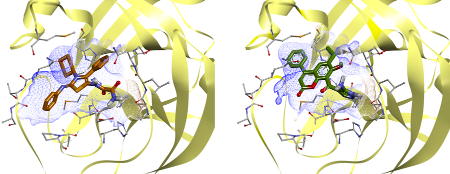 Example of complexes of ligands with main protease, obtained by molecular docking