
MitoMatrixCaL
Mitochondrial Calcium Sensor
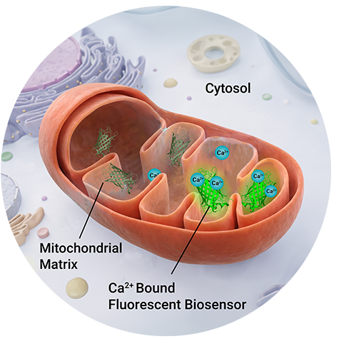
Mitochondria Calcium Localization; MitoMatrixCaL
Mitochondrial Ca2+ handling sits at the center of some of the most fundamental operations in cell biology including energy metabolism, apoptosis, reactive oxygen species (ROS) production, and cell signaling. Each pathway is affected by, and influences how mitochondria take up, buffer, and release calcium. Mitochondrial Ca2+ handling is central to many disease pathways and the Mitochondria Calcium sensor (MitoMatrixCaL) is built to enable researchers to examine Ca2+ dynamics in real time. MitoMatrixCaL lets you visualize mitochondrial Ca2+ uptake kinetics in response to physiological or pharmacological stimuli over time.
- Genetically encoded — Can be expressed in many in cell types, including primary neurons, iPSC-derived cell types, and immortalized cell lines; no exogenous dye loading or washout steps
- BacMam delivery — fast, efficient, dose-controlled transduction across a broad range of mammalian cell types, including cells that are difficult to transfect
- Live-cell, real-time — track dynamics from seconds or hours to days, rather than a single timepoint in fixed cells
- Mitochondrial matrix–targeted — a COX8 targeting sequence delivers the calcium sensor specifically to the mitochondrial matrix, monitoring a Ca2+ pool distinct from cytosolic or ER Ca2+ pools
- Built-in nuclear marker — no need to co-transduce a separate reagent for cell identification, segmentation or counting
- Screen in 384 well format or resolve single mitochondria— Same virus, same cells, no reoptimization. Plate reader and microscope compatible.
The mitochondrial and cytosolic Ca2+ responses to carbachol are distinct on a fluorescent plate reader
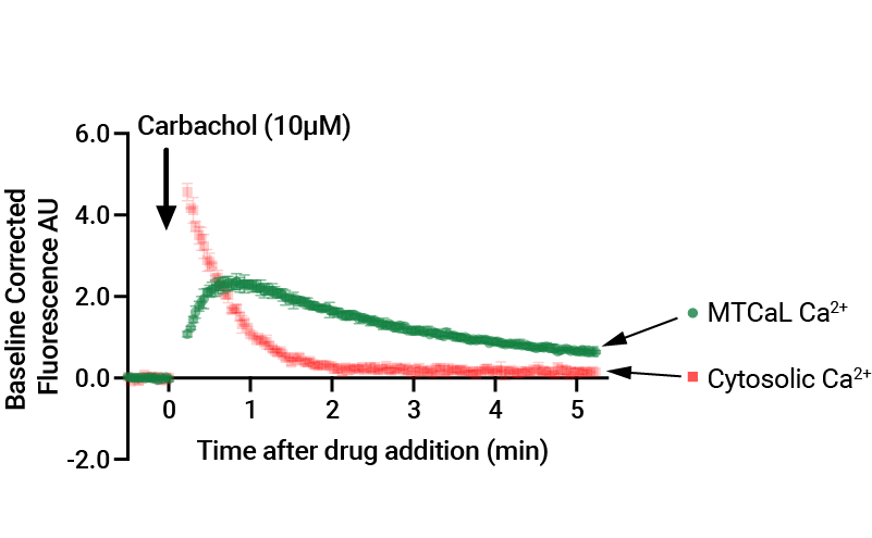
MitoMatrixCaL sensor was delayed in comparison to the cytosolic Ca2+ sensor (in red), which was expected. The initial increase in Ca2+ shown by the cytosolic Ca2+ sensor is due to the addition of carbachol activating muscarinic subtypes which evoke Ca2+ release. Mitochondria then take-up Ca2+ freely, as reported by the MitoMatrixCaL sensor. HEK293 cells were transduced with MTCaL or R-GECO (cytosolic Ca2+ sensor) and HM1 and stimulated with carbachol or vehicle control (not shown). Data was collected using a Synergy MX standard plate reader.
One reagent that scales from screening using a standard plate reader to looking at individual mitochondrion
Works on both microscope and plate readers

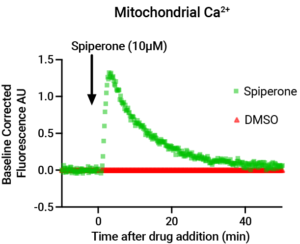
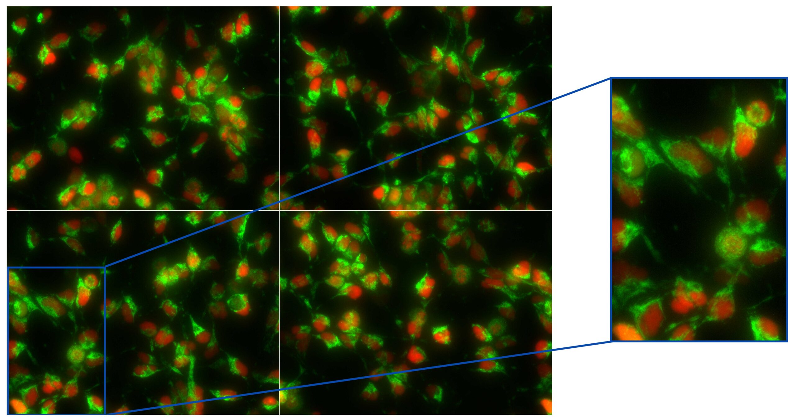
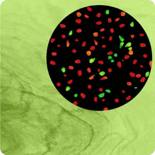
The MitoMatrixCaL delivers both a nuclear red marker and a green mitochondrial calcium sensor in a single BacMam construct. The addition of DMSO (left panel) does not activate the release of calcium. The addition of Spiperone increases mitochondrial Ca2+, likely via Ca2+ release first from the ER to the cytosol, then to the mitochondria (right panel). HEK293 cells were transduced with MTCaL and images were acquired on an EVOS M7000 imaging system.
This Spiperone response was easily read using a standard Synergy MX plate reader
Videos
Time lapse imaging of mitochondrial Ca2+ in AD293 Cells. MitoMatrixCaL delivers a red nuclear marker and a mitochondria matrix-targeted mNeonGreen Ca2+ sensor from a single BacMam construct. Both cell identification and mitochondrial Ca2+ can be tracked in the same field. AD293 cells were transduced with MitoMatrixCaL and imaged on an EVOS M7000 at 4 minute intervals over five hours. Small molecules that induce an increase in mitochondrial Ca2+ were added at t = 40 minutes. The timescale is shown in the upper left corner of each video
Vehicle (DMSO) left, and spiperone, 10µM right.
Vehicle (DMSO) left, and spiperone, 10µM right. The addition of spiperone raises cytosolic Ca2+ through a phospholipase C-dependent ER Ca2+ release (Antona et al. 2022) leading to mitochondrial uptake.
Vehicle (DMSO), left, and niclosamide, 10µM, right
Niclosamide impairs the growth of P53-deficient cells (tumor cells) but was originally developed for the treatment of intestinal tapeworm infections. Niclosamide is a hydrogen ionophore that translocates protons across the mitochondrial membrane inducing mitochondrial uncoupling (Kumar et al. 2018).
Vehicle (DMSO), left, and FCCP, 10µM, right
FCCP is a mitochondrial uncoupler that disrupts ATP production by transporting protons across mitochondrial membranes (Benz and McLaughlin 1983).
Mitochondrial Ca2+
Simultaneous measurement of mitochondria Ca2+ and cell motility in response to small molecule treatment. AD293 were transduced with MitoMatrixCaL and imaged on the EVOS M7000 every 4 minutes for several hours. Compounds were added at t=0: DMSO (Vehicle) spiperone (10µM), niclosamide (10µM) and FCCP (10µM).

Top Row: Mitochondrial Ca2+ reported by MitoMatrixCaL mNeonGreen Ca2+ sensor from segmented cells using FIJI.
Cell Motility

Bottom Row: Cell motility was quantified in FIJI as the frame to frame displacement of the mean position of the H2B red nuclear marker across the entire imaging field, expressed relative to vehicle control. On this scale, 0 indicates vehicle matched movement, and -1 indicates no detectable cell movement. Spiperone evoked a rapid transient rise in mitochondrial Ca2+ that peaked within 4 minutes and decayed to a sustained plateau, coupled with a modest reduction in movement. Niclosamide produced a small, transient increase in the mitochondrial Ca2+ signal that returned to baseline, together with a rapid loss of movement that reached the detection floor within 30 minutes and persisted for the remainder of the recording. FCCP produced a larger rise in mitochondrial Ca2+ signal followed by a sustained plateau above baseline, and a comparably rapid and sustained loss of movement.

TRPML1-Lysosomal-Ca2+ sensor (TLC) for the Ca2+ Dyshomeostasis Neurodegeneration Model
Here we present a genetically encoded TRPML1 Lysosomal Ca2+ (TLC) sensor that enables direct measurement of Ca2+ release with high specificity. Using this tool we show that small molecule agonists differ in their ability to selectively activate TRPML1, with MSK-83 demonstrating higher specificity compared to ML-SA5. The Assay provides a powerful approach for interrogating TRPML1 function in human cells and for identifying modulators of lysosomal Ca2+ signaling.
Perturbed Ca2+ signaling leads to synaptic loss, amyloid and tau aggregation, mitochondrial dysfunction, lysosomal alkalinization, and superoxide production, all of which are hallmarks of neurodegenerative diseases. Evidence from Alzheimers models suggest that ER-derived Ca2+ signals disrupt mitochondria-lysosome interactions, further exacerbating cellular stress.
GPCR Biology
Targeted GECO Calcium Assay Kits
Showing all 2 results

 Calcium Assays, GPCR Assays, Targeted GECO Calcium
MTCal-Mitochondria-Targeted G-GECO Ca2+ Assay Kit – Big Sky (U0652G)
$995.00Select options This product has multiple variants. The options may be chosen on the product page Calcium Assays, GPCR Assays, Targeted GECO Calcium
TRPML1-Targeted G-GECO Ca2+ Assay Kit – Big Sky (U0653G)
Read more
Shop Other Assay Kits by Category

Products
GPCRs in BacMam Virus
Targeted GECO Ca2+
mNeonGreen BacMam Tools
Product U0652G
Products U0221R & D0221G
Product U0654G
Product P1040G
Product P1010G
MitoPaint COX8
Products U0261G , U0262G, U0267G, U0266G
Products U0241R, D0241G, U0267G, D0231G
Product D0331G
CAAX