Express your neurodegenerative-associated protein of interest with bright fluorescent biosensors for cell stress and signaling in your favorite neuronal culture or cell line.
See how to capture neurodegenerative processes in living cells.
”Live-cell assays for cell stress responses reveal new patterns of cell signaling”
Identify cell stress from misfolded proteins in the ER and cytoplasm
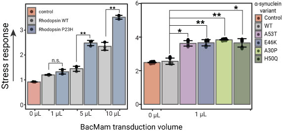
n.s. = not significant, * = pval < 0.05, ** = pval < 0.01
Detection of cell stress using the ratiometric cell stress biosensor (#U0901G).
Left: Stress detection in cells expressing WT and mutant rhodopsin, which folds in the ER.
Right: Stress detection of cells expressing WT and mutant versions of α-synuclein, which folds in the cytoplasm.
Compare cell signaling responses in cells expressing WT or mutant proteins

HEK293T cells expressing the R-GECO Ca2+ biosensor (#U0600R) and WT or mutant P23H rhodopsin were stimulated with carbachol to produce a Gq-dependent Ca2+ signaling response. Mutant rhodopsin expression alters the Ca2+ signaling response
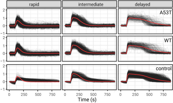
Use optogenetic tools to examine cell signaling in neurodegenerative models
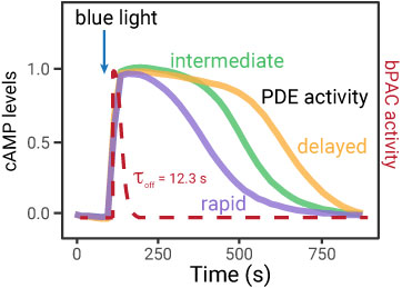
PDE activity profiles in cells expressing WT and A53T mutation α-synuclein protein.